Wednesday, December 26, 2012
Tuesday, December 25, 2012
A prospective study in detection of lower-limb lymphedema and evaluation of quality of life after vulvar cancer surgery.
Jul 2012
Source
Department of Obstetrics and Gynaecology, 2nd Medical Faculty of Charles University in Prague, Czech Republic. marta.novackova@seznam.cz
Abstract
BACKGROUND:
Lower-limb lymphedema is one of the most disabling adverse effects of vulvar cancer surgery. Multifrequency Bioelectrical Impedance Analysis (MFBIA) is a modern noninvasive method to detect lymphedema. The first aim of this study was to prospectively determine the prevalence of secondary lower-limb lymphedema after surgical treatment for vulvar cancer using objective methods, circumference measurements and MFBIA technique. The second aim was to compare quality of life (QoL) before and 6 months after vulvar surgery.
METHODS:
Twenty-nine patients underwent vulvar cancer surgery in our study: 17 underwent inguinofemoral lymphadenectomy (RAD), and 12 underwent sentinel lymph node biopsy (CONS). Patients were examined before and 6 months after vulvar surgery by measuring the circumference of the lower limbs and with MFBIA. A control group of 27 healthy women was also measured. To evaluate QoL, the European Organisation for Research and Treatment of Cancer (EORTC) QoL questionnaires (QLQ-C30 and QLQ-CX24) were administered to patients before and 6 months after surgery.
RESULTS:
Using circumference measurement, 9 lymphedemas (31%) were diagnosed: 3 (25%) in the CONS and 6 (37.5%) in the RAD group (P = 0.69). After vulvar surgery, patients in the RAD group reported more fatigue and worsening of physical and role functioning. When comparing both groups, the RAD group had significantly worse parameters in social functioning, fatigue, and dyspnea.
CONCLUSIONS:
Lower radicality in inguinofemoral lymphadenectomy shows a trend toward lower morbidity and significantly improves QoL. Multifrequency Bioelectrical Impedance Analysis was tested in these patients as a noninvasive, objective method for lymphedema detection. Detection of lymphedema based on subjective evaluations proved to have an unsatisfactory sensitivity. Less radical surgery showed objectively better results in QoL.
Distichiasis-lymphedema syndrome with optic disc pit
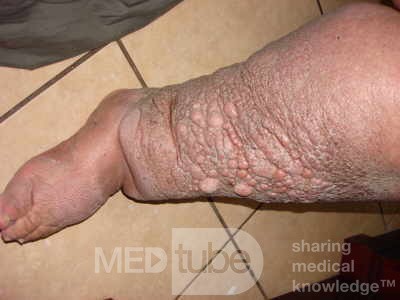
Leg Lymphedema
Jan-Feb 2011
Indian J Ophthalmol. 2011 Jan-Feb; 59(1): 71–72.
Dear Editor,
An eight-year old boy, first born to third degree consanguineous parents, presented with right leg swelling for three months, with gradual onset, which progressed up to knee. There was no history of fever, injury, abdominal pain or contact with tuberculosis. He was treated with anti-filarial drugs elsewhere. At two years of age, he had frequent episodes of redness and constant rubbing of eyes and was then diagnosed to have double-rowed eye lashes involving all four eyelids and the extra rows of lashes were cauterized and removed elsewhere. The boy still continued to be symptomatic. None of the other family members had similar complaints.
On examination, he had right lower limb edema, which was from the knee downward. There were no bony deformities or vertebral anomalies. Systemic examination was normal. He had mild congestion of both eyes. His visual acuity was 20/20; N6 in both eyes, and had no refractory error. Slit-lamp examination revealed distichiasis. focal area of loss of eyelashes and depigmentation of skin was noted in the left upper eyelid. Fundus examination revealed an optic disc pit in the left eye and the macula was normal
Blood parameters were normal. Night smears for microfilaria were negative. Ultrasonography (USG) abdomen, echocardiogram, magnetic resonance imaging (MRI) spine, and vascular Doppler studies of both limbs were normal. Isotope lymphoscintigraphy confirmed the lymphedema. The parents were also screened and found to be normal. A clinical diagnosis of distichiasis-lymphedema syndrome (DLS) was made. Conservative management for symptomatic distichiasis, with lubrication and epilation was carried out, advice for Amsler test at home periodically and stockings for lymphedema were given. The parents were genetically counseled for prevention of secondary complications such as, cellulitis, foot infections, and varicose veins.
Full Text Article:
Thursday, December 20, 2012
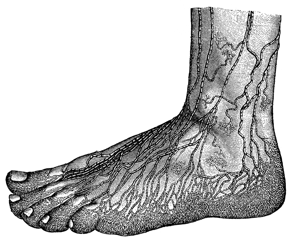
Lymph nodes of the foot
Lymph supply of the feet
Bellissimabeauty
Lymph supply of the feet
3d4 Medical
ClipArt
See also:
Lymph nodes
Lymph supply of the feet
Bellissimabeauty
Lymph supply of the feet
3d4 Medical
ClipArt
See also:
Lymph nodes
Labels:
feet,
foot,
lymph node,
lymph supply,
lymphedema
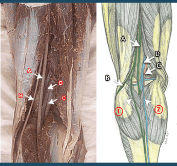
Popliteal lymph nodes
Popliteal lymph nodes - located in the knee area
The small popliteal lymph nodes are four or five in number and surround the popliteal veins and arteries. They are clustered at the back part of the leg behind the knee joint. They help collect excess fluids from your feet and legs.
Organatomy
King Saud University
Dartmouth
The small popliteal lymph nodes are four or five in number and surround the popliteal veins and arteries. They are clustered at the back part of the leg behind the knee joint. They help collect excess fluids from your feet and legs.
- Popliteal artery and its branches
- Popliteal vein and its tributaries
- Tibial and common peroneal nerves.
- Termination of the small saphenous vein.
- Lower part of the posterior cutaneous nerve of the thigh.
- Popliteal lymph nodes, connective tissue and fat.
Organatomy
King Saud University
Dartmouth
Leg and Inguinal Lymph Nodes
The inguinal nodes - groin area. I had a lymphangiogram done in 1966 and a lymphoscintigraphy done in 2006 - and they showed me missing significant numbers of inguinal nodes. In 1962, when I was nine years old, I had an inguinal node removed due to necrosis from cellulitis - in left leg.
In 2000, I had a small needle biopsy on an inguinal node in the right leg. Yep....it was positive.
In 1995, I was diagnosed with mixed b-cell lymphoma.
Wellsphere
Plymouth Hospitals
Lymphedema People
Lymph nodes - female. Though the abdominal nodes will be different due to different organs, the inguinal nodes remain the same.
Healthtap
Hopkins
Thursday, December 13, 2012
Lymphoscintigraphy in unilateral lower limb and scrotal lymphedema caused by filariasis.
Dec 2012
Source
Department of Nuclear Medicine and PETCT, Amrita Institute of Medical Sciences, Cochin, Kerala, India.
Abstract
Lymphedema is the edema that results from chronic lymphatic insufficiency. Lymphatic filariasis is caused by the filarial nematodes Wuchereria bancrofti, Brugia malayi, and Brugia timori. Lymphatic filariasis is common in tropical and subtropical regions. Early diagnosis and prompt therapy can be implemented using lymphoscintigraphy. Our patient is a 15-year-old boy presenting with a 3-month history of hydrocele. The patient was referred to us to rule out any lower limb lymphatic obstruction as the patient is from an endemic area. Tc Sulfur colloid (filtered) lymphoscintigraphy showed abnormal tracer collection in the scrotum and penis. There is associated dermal backflow or stasis in the left thigh region extending just above the knee, suggesting partial obstruction of left inguinal lymphatic channels.
See also:
Sunday, December 2, 2012
How to Bandage Wrap the Lymphedema Leg
One of the best posts on how to wrap a leg…from one of my online members:
Since you have the swelling in the feet (and toes), it is probably lymphedema, perhaps compounded with lipedema. The traditional bandaging technique is with a stockinet, then some artiflex (cotton padding), and lastly, the bandages. I bandage directly over the skin. The padding is supposed to even out if you should constrict some part of the bandaging, causing the lymph not to flow, but the bandages are really not like rubber bands – properly spaced and overlapped, they will not cause constriction – and the artiflex is a pain. The stockinet is just another thing to wash and dry. I went to a bandage supplier (now out of business) and found that they have new bandages that are thick enough to be used without layering (e.g. the stockinet and padding).
Perhaps this is the way to go, or perhaps you want to bother with stockinets and padding. If you were seeing a therapist, they would also use foam instead of artiflex (just cotton padding). Some pictures of bandaging look absolutely monstrous. My so called therapist used some foam, etc., but I soon discovered that the leg went down more without it. The pad is supposed to “spread” the compression so there is no binding – but what really happens is all the elasticity of the bandages goes to compressing the FOAM – not compressing your leg. A little compression trickles down to the actual leg, but my experience was that the swelling went down better without the extra stuff. However, since this is against tradition, you should at least be aware if any part of your leg feels too tight, and, if so redo the bandages (which is at least an hour for two legs – and bandages that were OK while you were up and around can suddenly become too tight in the middle of the night – which means you have to get up and do it again.)
Anyway, with or without stockinet and padding, here is one technique for bandaging:
Materials (1 large leg not grossly larger than normal (I am 5'9” and the calf measure is 21” and I have wide, swollen feet - if you are substantially larger, you may need more)
optional: stockinet, artiflex, foam
required:
1 roll 1” professional strength masking tape. 1 ea 3” strip of heavy padding around the ankles 1 ea 1” x 5m medi-rip 2 ea 8 cm. x 5 m short stretch bandages 1 ea 10 cm x 10 m short stretch bandages 1 ea 6 cm x 5 m short stretch bandages.
Double for 2 legs, if you are very much larger than me, add another 1 ea 10 cm. x 5 m short stretch bandage for each leg.
I sit on my bed and have a low table I can rest my foot on, but two chairs will work also (one to sit on and one to put your foot on).
Wrap the 3” strip of heavy padding (or chock pads) around the ankles. The figure 8's you are making around your foot and from the foot onto the leg will tend to bind right at the intersection of the foot and leg (where the 90” turn is made. This is the only place padding is essential.
Secure it with masking tape. Secure all the bandages after they have been wrapped with masking tape. Cut a lot of 5” strips of masking tape and have them ready. Stick them on the edge of the table, or a windowsill, or something.
First hold all the bandages so that you are drawing from the bottom of the bandage cylinder (the bandages rolled up are a cylinder), not the top. A little experimentation will show you that this is much easier.
Start with the 1” medi-rip (it is a self cohesive bandage, but looses some of the self cohesion with laundering). Use this tiny bandage to bandage along the toe line. That is, make the same arc that the joints of the toes to the feet make. Do not bind the toes. If you can, wrap each toe with it, but I find that this binds the toes and hurts, so I leave my toes unwrapped, even though they swell, but if you start with the larger short stretch bandages, there will be a half moon that swells even more (Since if you make a straight circle from just below the little toe to just below the big toe, this will leave some area of foot not bandaged and the lymph will be pushed into this area, and it will be worse than before. The little 1” medirip can be wrapped in a curved path that covers all of the foot. Overlap this 1” medirep by 1/2 and continue winding it around your foot until you get to the end of the arch, then take it up diagonally over the top of the foot, and you will still have enough bandage to wrap again just under the toe line again for a few wraps. The medi wrap has strands of elastic in an otherwise cotton strip, so pull the medirip tight (that is the elastic is extended, but not to the point of discomfort).
When you wrap the bandages, pull a bit at the end of each circle, but do not stretch them too hard, or with constant tension as far as they will stretch. You want them to exert a little spring, but don't strangle your legs. If you get them too tight, it will hurt, and you must undo your wrapping and redo it (a big pain). If you don't stretch them a little, they won't have much compression. Of course, it's always the bottom bandages on the feet that hurt, so you have to unwrap the whole deal to get to them.
Next,step 2 take a 8 cm. x 5 m short stretch bandage, and start at the tip of the foot, but do not bind any toes, and since you already have the medi-rip, allow a little breathing space to make sure you don't bind toes. Then wind around your foot overlapping the bandages by about 1/2 to 2/3 (I probably overlap 2/3) until you have gotten almost to the leg (your foot should be at a 90 degree angle to the leg, and for me this is 2 or 3 wraps), then go around the heel itself, and, as you come off the other side of the heel, take the bandage diagonally up on the top of the foot to just below the top of the first wrap (just under the bottom of the big toe), go around the bottom of the foot, and then bring the bandage back around the ankle just above the heel, then around the ankle, and back up diagonally across the top of the foot just like before, overlapping 1/2 to 2/3 of the previous path. This will make large figure 8s.
Continue with the figure 8's each layer a little higher around the ankle, until you again are wrapping just in front of the leg (no more space to do another figure 8) and use the rest of the bandages going in straight circles (not figure 8's) around the ankles.
Next,step 3 take the second 8 cm x 5 meter short stretch bandage, and start at the base of the leg (around the ankles), go around once or twice, to anchor the bandage, then on the next turn go down around the bottom of the foot close to the heel, and then around the bottom of the foot and then over and up around the leg, then continue making figure 8's up the leg overlapping by about 2/3.
To make a figure 8 around the leg, on one side of the front of the leg, the bandage is going uphill (or towards your knee), then it goes more or less straight around the back of the leg at the high end of the 8, then goes downhill (or towards the foot), as you come across the front of the leg again, then more or less straight across the back of the leg at the low end of the 8 and then up again for the next figure 8. On me, this bandage is finished just about at the beginning of the calf (a little above the bottom of the muscle – it would be ideal if this bandage ended just before the muscle begins, but it will be a bit different for everyone depending of how much they overlap and how large their leg is.
Next,step 4 do figure 8's with the 10 cm x 10 m bandage. Begin at the bottom of the leg with the beginning of the bandage facing upward, so the first direction is in a downward direction, (the end pointing up) coming around and then going up again. The 10 cm x 10 m bandage should take you up to just below the knee, but if the legs are very large, you may need another 10 cm. bandage. Each course of the figure 8 should overlap a little less or evenly, but not more than the previous course. The more you overlap the greater the compression, and you must always have less compression proximally (towards your heart) than distally (towards your toes).
Finally,step 5 take the last 6 cm. x 5 meter short stretch bandage and start at about mid calf or a little higher, and wind in straight circles until just below and as close as possible to the knee. This last bandage gives compression over the tops of the top 8's where there is not as much overlap, and sort of holds it all up, as the circumference of the leg is actually smaller at the knee than at the mid calf (doesn't slide down because a smaller circle would have to slide over a larger circumference of the leg).
I have been complemented on my ability to wrap, but It is hard to know if a novice can make much sense of my directions – but I tried. You don't see to many photographs of the figure 8's, but they give more compression and stay up better, and bind less. You will get the general idea of winding up the leg, and overlap by looking at the photographs, however. It may seem complicated to follow my directions (I tried to be clear), but the real technique is not very hard at all.
The new thick bandages that do not need padding (padding is included) are : KomprimED. They are located on the bandagesplus web site under bandages, then under two way stretch bandages. I think you should start with these, as the padding may be more important for someone who is just beginning bandages. This is much simpler than all those stupid layers.
*Soft and comfortable directly on patient's skin *Thicker texture avoids application of foam and padding in many cases *Suitable for lymphedema and venous ulcers *Patient-friendly application requires less layers *All bandages are short-stretch/low stretch KomprimED 4cmx5m
Other wise, the standard short stretch bandages are rosidal or comprilan. I use rosidal. The medi-rip is under the section cohesive bandages on page 2 under the more general category bandages.
Suppliers of Bandages, Wrapping materials and Supplies:
Labels:
bandage wrap,
leg lymhpedema,
materials,
short stretch bandage
Sunday, November 25, 2012
Secondary Leg Lymphedema
Introduction
If you ask most people that are familiar with lymphedema the question, “Are you aware of secondary lymphedema,” most would reply that “yes, it is where the arm swells after the lymph system has been damaged by breast cancerbiopsy and treatment.” This is called arm lymphedema.
Even if they are aware that such a condition as secondary leg lymphedema exists, their response might well be that it is a small group of afflicted men who have prostate cancer.
Thus shows how little awareness there is about this particular form of lymphedema. Even in the lymphedema world it is a poor step-child.
However, if the membership of Lymphedema People and the posts in the online lymphedema support groups are an indication, this condition is increasing dramatically.
The reasons for this increase are multiple. They include:
1. increased survival rates of cancer 2. improved treatment of trauma injuries that previously would have been terminal 3. increase in antibiotics for infections and treatment for other conditions that previously might have resulted in death.
It is also important to note that secondary leg lymphedema does not necessarily start immediately after the injury or trauma. It may not start for years.
What is secondary leg lymphedema?
Secondary lymphedema is a condition where the lymphatic system has been damaged. The main job of this system is to move excess through and out of our bodies. When it becomes damaged or impaired, it is no longer able to accomplish this function and these fluids (lymph fluids) collect in the interstitial tissues of our legs. This causes leg swelling.
Another important function of the lymph system is to help our bodies fight infections. With lymphedema, this ability is also weakened and the patient becomes more susceptible to infections.
What causes secondary leg lymphedema?
Secondary leg lymphedema (also referred to as acquired lymphedema) is caused by or can develop as a results of:
1.) Surgeries involving the abdomen or legs where the lymph system has been damaged. This includes any intrusive surgery.
Examples are
vein stripping surgery for peripheral vascular disease hip replacement knee replaement insertion of bolts, screws and other devices in orthopaedic repair lipectomy
2.) Removal of lymph nodes for cancer biopsy. These cancers include, but are not limited to
prostate cancer testicular cancer ovarian cancer uteran cancer vulva cancer bladder lymphoma - both hodgkins and non hodgkinsmelanoma colon Kaposi Sarcoma
3.) Radiation treatment of these cancers that scars the lymph system and lymph nodes
4. Some types of chemo therapy. For example, tamoxifen has been linked to secondary lymphedema and blood clots.
5.) Severe infections/sepsis. Generally referred to as lymphangitis, this is a serious life-threatening infection of the lymph system/nodes.
6.) Trauma injuries such as those experienced in an automobile accident that severly injures the leg and the lymph system.
7.) Burns - this even includes severe sunburn. We have a member that acquired secondary leg lymphedema from this.
8.) Bone breaks and fractures.
9.) Morbid obesity - the lymphatics are eventually crushed by the excessive weight. When that occurs, the damage is permanent and chronic secondary leg lymphedema begins.
10.)Insect bites
What are some of the symptoms of secondary leg lymphedema?
These symptoms may include:
1.) Unexplained swelling of either part of or the entire leg. In early stage lymphedema, this swelling will actually do down during the night and/or periods of rest, causing the patient to think it is just a passing thing and ignore it.
2.) A feeling of heaviness or tightness in the leg
3.) Increaseing restriction on the range of motion for the leg.
4.) Unsual or unexplained aching or discomfort in the leg.
5.) Any change involving hardening and/or thickening of the skin or areas of skin on the leg.
Labels:
causes,
edema,
infections,
injury,
lymphatics,
pain,
risk factors,
secondary leg lymphedema,
skin,
swelling,
symptoms,
trauma
Wednesday, November 14, 2012
Congenital Lymphedema : Another Unique and Gender Specific Stigmata of Tuberous Sclerosis?
Congenital lymphedema : another unique and gender specific stigmata of tuberous sclerosis?
Nov 2012
Source
Department of Cardiology, SCTIMST, Thiruvananthapuram, Kerala, India.
We report a child of tuberous sclerosis with a rare association of congenital lymphedema and cardiac rhabdomyoma since birth.
A 3-month-old female child, born of non-consanguineous marriage , was detected soon after birth to have nonpitting edema of left lower limb extending from thigh to foot. Neurosonogram and USG abdomen were normal. Echocardiography revealed a 9 x 9 mm rounded pedunculated mass in LV outflow tract, attached to aorto-mitral continuity junction. Physical examination revealed multiple hypopigmented macules in right upper limb and trunk suggestive of ash leaf macules. X-ray chest and ECG was normal for age. Blood investigations were normal. The child’s father had a history of seizure disorder and was on antiepileptic drugs. His physical examination revealed hypopigmented to depigmented macules in both upper limbs and trunk, skin colored plaques with irregular border in lumbosacral region and multiple hyperpigmented to erythematous papules and small plaques over face suggestive of ash leaf macules, shagreen patches, and angiofibromas respectively.
Lymphedema is a chronic tissue swelling that is most commonly manifested in a limb. This condition results from impaired lymph drainage in the presence of normal capillary filtration. The three main consequences of lymphatic failure are lymphedema, infection and, very rarely, cancer [1]. Most forms of primary lymphedema are thought to be caused by a congenital abnormality of the lymphatic system and present at or soon after birth. Cardiac rhabdomyomas are intracavitary or intramural tumors that are present in nearly 50 to 70% of infants with tuberous sclerosis (TSC). Most children are asymptomatic. Symptoms are attributed to the presence of intracardiac obstruction, myocardial involvement, and rhythm disturbances [2].
Congenital lymphedema is a rare association with tuberous sclerosis with only few cases reported earlier [3,4]. The previous reported patients were females but unlike our child, they presented with history of multiple seizures while our child had no seizures but instead had a cardiac rhabdomyoma detected incidentally. It is interesting to note that pulmonary lymphangio-myomatosis seen in tuberous sclerosis similarly occurs only in women which is hypothesized to be due to the fact that estrogen regulates TSC gene signalling and, perhaps, also the migration of TSC2-deficient cells [3].
The pathophysiology of congenital lymphedema in tuberous sclerosis is yet unclear. Previous authors have suggested that it could be due to the dysplastic development of lymphatic system in the affected limb as part of TSC gene mutation as this gene regulates cell growth, proliferation and migration. Congenital lymphedema may also be due to the abnormal smooth cell hypertrophy in subcutaneous tissue which externally compresses the superficial lymphatics. An increased awareness of this association may help pediatricians suspect tuberous sclerosis in a female child when congenital lymphedema is the sole external manifestation.
Friday, November 9, 2012
A newly designed SIPC device for management of lymphoedema.
WARNING TO ALL LYMPHEDEMA PATIENTS
This abstract is a prime example of just how bad information can be that is presented even through PubMed.
Lymphology 101 clearly shows that high pressure pneumatic devices can cause serious damage to the good lymphatics, making lymphedema even worse.
Read this for education, but please, please, please, what ever you do never ever ever set the compression level on high if you use a pneumatic device.
A newly designed SIPC device for management of lymphoedema.
PubMed
Pat
Labels:
edema,
lymphatic system,
lymphedema,
pnuematic device,
treatment,
warning
Wednesday, November 7, 2012
Lambda-shaped anastomosis with intravascular stenting method for safe and effective lymphaticovenular anastomosis.
2011
Source
Department of Plastic and Reconstructive Surgery, Graduate School of Medicine, University of Tokyo, Tokyo, Japan. tyamamoto-tky@umin.ac.jp
Abstract
BACKGROUND:
Lymphaticovenular anastomosis has become an increasingly common treatment for lymphedema. Supermicrosurgical techniques are essential for the successful performance of lymphaticovenular anastomosis. A positive correlation between the number of lymphaticovenular anastomoses performed and therapeutic efficacy has been reported, and in performing these anastomoses, the establishment of as many bypasses as possible is important.
METHODS: Forty limbs of 20 patients with lower extremity lymphedema who underwent lymphaticovenular anastomosis in our department were assessed. All cases were performed under local anesthesia using two to four surgical microscopes. A new method of anastomosis, lambda-shaped anastomosis assisted by intravascular stenting, was chosen in required cases.
RESULTS: Lymphaticovenular anastomoses resulted in 186 anastomoses on 20 patients with lower extremity lymphedema; the average number of anastomoses per case was 9.3 (range, five to 18). The number of surgical microscopes used ranged from two to four (average, 3.3), and the duration of the operation ranged from 3 to 5 hours (average, 4.1). In the cases of lambda-shaped anastomosis (n = 11), the number of anastomoses was significantly greater than in the cases without lambda-shaped anastomosis (n = 9; 10.2 ± 2.3 versus 8.2 ± 1.4; p < 0.05).
CONCLUSIONS:
Lambda-shaped anastomosis assisted by intravascular stenting is a safe and relatively easy method that can be performed by surgeons with less than 1 year of experience in microsurgery. This in turn allows efficient lymphaticovenular anastomoses to be performed simultaneously by a team of surgeons, resulting in an increased number of bypasses.
Lippincott, Williams & Wilkins
*Editor's Note: Article is for information only and is NOT to be construed as an endorsement of the procedure, nor as a position of opposing the procedure.
Subscribe to:
Posts (Atom)